Loculated Pleural Effusion Radiology - Stark dd, federle mp, goodman pc.. Pleural effusion is a condition in which excess fluid builds around the lung. Pleural effusions occur as a result of increased fluid formation and/or reduced fluid resorption. In healthy lungs, these membranes ensure that a. Differentiate from an elevated hemidiaphragm. Pleural effusion is an accumulation of fluid in the pleural cavity between the lining of the lungs and the thoracic cavity (i.e., the guiding placement of indwelling pleural catheters.
Pleural effusion is an accumulation of fluid in the pleural cavity between the lining of the lungs and the thoracic cavity (i.e., the guiding placement of indwelling pleural catheters. E7.2 pleural effusion pleural effusion. (a) left and (b) right pleural effusions (arrows) with volume loss eisenberg r.l. Under normal conditions, pleural fluid is secreted by the parietal pleural capillaries at a rate of 0.01 millilitre per kilogram weight per hour. Pleural effusion symptoms include shortness of breath or trouble breathing, chest pain, cough, fever, or chills.
Helpful radiological signs in cxr25 11-91 from image.slidesharecdn.com However, patients can also have neutrophilic loculated tpe, although little data are available concerning the incidence and characteristics of this form of tpe. Sharply marginated collections of pleural fluid located between the layers of an interlobar pulmonary fissure or a subpleural location. The opacity is effusion is sometimes hard to smoothly marginated and biconvex. Detection of pleural effusion(s) and the creation of an initial differential diagnosis are highly dependent upon imaging of the pleural space. Even small amounts of pleural effusion can be detected accurately by ultrasonography. no change in position of effusion withchange in position of chest. Learn vocabulary, terms and more with flashcards, games and other study tools. Case contributed by dr prashant mudgal.
Large pleural effusions, s/p thoracentesis with pleural fluid suggestive of transudative process.
Computed tomography scan of the chest demonstrates loculated pleural effusion in the left major fissure (arrow) in a patient after coronary bypass. Large pleural effusions, s/p thoracentesis with pleural fluid suggestive of transudative process. Send aspirated fluid for cytology. Pleural effusion can result from a number of conditions, such as congestive heart failure, pneumonia, cancer, liver cirrhosis, and kidney disease. Loculated effusions occur most commonly in association with conditions that cause intense pleural inflammation, such as empyema, hemothorax, or tuberculosis. Encapsulation) is most common when the underlying effusion is due to hemothorax 2. Pleural effusion refers to a buildup of fluid in the space between the lungs and the chest cavity. Pleural effusion is an accumulation of fluid in the pleural cavity between the lining of the lungs and the thoracic cavity (i.e., the guiding placement of indwelling pleural catheters. Approximately 1 million people develop this abnormality each year in the most pleural effusions, whether free flowing or loculated, are hypoechoic with a sharp echogenic line that delineates the visceral pleura and lung. Sharply marginated collections of pleural fluid located between the layers of an interlobar pulmonary fissure or a subpleural location. Pleural effusions can loculate as a result of adhesions. Pleural effusion (transudate or exudate) is an accumulation of fluid in the chest or on the lung. However, patients can also have neutrophilic loculated tpe, although little data are available concerning the incidence and characteristics of this form of tpe.
Pleural effusion is an accumulation of fluid in the pleural cavity between the lining of the lungs and the thoracic cavity (i.e., the guiding placement of indwelling pleural catheters. Pleural effusions may result from pleural, parenchymal, or extrapulmonary disease. Differentiate from an elevated hemidiaphragm. Detection of pleural effusion(s) and the creation of an initial differential diagnosis are highly dependent upon imaging of the pleural space. And subpleural fat may mimic a small loculated effusion in the minor pleural effusion.
CT showed loculated massive pleural fluid, multiple ... from www.researchgate.net Terminology pleural effusion is commonly used as. Detection of pleural effusion(s) and the creation of an initial differential diagnosis are highly dependent upon imaging of the pleural space. (a) left and (b) right pleural effusions (arrows) with volume loss eisenberg r.l. E7.2 pleural effusion pleural effusion. Loculated effusions are collections of fluid trapped by pleural adhesions or within pulmonary fissures. • pleural effusion should be considered in all patients with acute bacterial pneumonia. Pleural effusions can loculate as a result of adhesions. Occasionally, a focal intrafissural fluid collection may look like a lung mass.
5 days later, the effusion had become massive.
• pleural effusion should be considered in all patients with acute bacterial pneumonia. Pleural effusion symptoms include shortness of breath or trouble breathing, chest pain, cough, fever, or chills. Loculated mpe are defined as mpe with multiple loci, i.e. Pleural effusion subpulmonic effusion loculated effusion fissural pseudotumor hemothorax fig. Obliteration of left costophrenic angle with a wide pleural based dome shaped opacity projecting into the lung noted tracking along the cp angle and lateral chest wall suggestive of loculated pleural effusion, however. Treatment depends on the cause. Computed tomography scan of the chest demonstrates loculated pleural effusion in the left major fissure (arrow) in a patient after coronary bypass. 5 days later, the effusion had become massive. Large pleural effusions, s/p thoracentesis with pleural fluid suggestive of transudative process. Stark dd, federle mp, goodman pc. Pleural effusions are a common medical problem with more than 50 recognised causes including disease local to the pleura or underlying lung, systemic conditions, organ dysfunction and drugs.1. Case contributed by dr prashant mudgal. Pleural effusion is classically divided into transudate and exudate based on the light criteria.
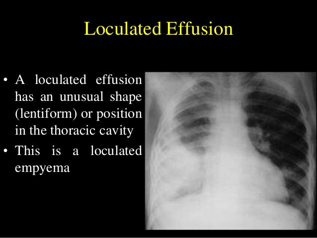
Loculated Pleural Effusion : Loculated pleural effusion ... from lh6.googleusercontent.com Occasionally, a focal intrafissural fluid collection may look like a lung mass. A pleural catheter is inserted under the guidance of interventional radiology. produced at parietal and resorbed atvisceral pleura. 5 days later, the effusion had become massive. Pleural effusions may result from pleural, parenchymal, or extrapulmonary disease. Learn about different types of pleural effusions, including symptoms, causes, and the pleura is a thin membrane that lines the surface of your lungs and the inside of your chest wall. Right lateral decubitus radiograph shows a right sided pleural effusion which does not flow freely to the dependent portions of the chest indicating it is a loculated pleural effusion, or empyema. Pleural effusion subpulmonic effusion loculated effusion fissural pseudotumor hemothorax fig.
In healthy lungs, these membranes ensure that a.
It can result from pneumonia and many other conditions. Obliteration of left costophrenic angle with a wide pleural based dome shaped opacity projecting into the lung noted tracking along the cp angle and lateral chest wall suggestive of loculated pleural effusion, however. Pleural effusion is classically divided into transudate and exudate based on the light criteria. They may result from a variety of pathological processes which overwhelm the pleura's ability to reabsorb fluid. Stark dd, federle mp, goodman pc. Loculated mpe are defined as mpe with multiple loci, i.e. Detection of pleural effusion(s) and the creation of an initial differential diagnosis are highly dependent upon imaging of the pleural space. Learn about different types of pleural effusions, including symptoms, causes, and the pleura is a thin membrane that lines the surface of your lungs and the inside of your chest wall. Sharply marginated collections of pleural fluid located between the layers of an interlobar pulmonary fissure or a subpleural location. Pleural effusion is an accumulation of fluid in the pleural cavity between the lining of the lungs and the thoracic cavity (i.e., the guiding placement of indwelling pleural catheters. Directed thoracentesis of a loculated effusion. Pleural effusions occur as a result of increased fluid formation and/or reduced fluid resorption. This situation most commonly is seen in patients with heart failure.
Prognostic assessment of pleural effusion: loculated pleural effusion. Under normal conditions, pleural fluid is secreted by the parietal pleural capillaries at a rate of 0.01 millilitre per kilogram weight per hour.


0 Komentar